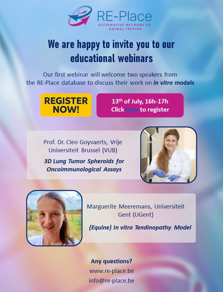
Kick-off Educational Webinars
In order to promote the use and development of New Approach Methodologies, we are organizing a series of Educational Webinars putting the existing knowledge in the spotlight!
Practical information
The Webinars are open to all interested parties including scientists, regulators, the authorities, and the public. During every session, two scientists will be given the opportunity to present their work to their peers. All webinars will be recorded and will be placed on the RE-Place YouTube Channel: https://www.youtube.com/@re-place1330
Participation is free of charge, but registration is mandatory via following: https://shorturl.at/djwNU
First session
The first session will take place on the 13th of July from 16h to 17h via Webex and will welcome two researchers from the RE-Place database who are developing in vitro models.
- Prof. Cléo Goyvaert (Vrije Universiteit Brussel) will talk about the development of 3D lung tumor spheroid platform. After obtaining a Bachelor degree in Veterinary Sciences and Master in Biochemistry and Biotechnology at University of Ghent, Cleo Goyvaerts started a PhD project in the field of antitumor vaccination at the Vrije Universiteit Brussel (VUB). As junior postdoc, she focussed on the manipulation of the tumor microenvironment, partly at Mount Sinai Icahn School of Medicine, New York. Since 2018, she works as Assistant Professor at VUB where she focusses on resistance mechanisms that hamper curative immunotherapy for lung cancer.
- Marguerite Meeremans (Universiteit Gent) will present the in vitro tendinopathy model that she is developing as part of her PhD thesis under the supervision of Prof. dr. Catharina De Schauwer at the Veterinary Stem Cell Research Unit of the Faculty of Veterinary Medicine. Her research focuses on the development of a physiologically representative tendon injury model by combining equine cells (tendon cells, endothelial cells, macrophages), chemically modified gelatin, 3D printing and mechanical stimulation. Once this model is established, underlying pathways of tendon pathogenesis and novel therapies, such as mesenchymal stem cells and their secretome, can be studied in a controlled and representative environment bypassing the confounding factors associated with in vivo clinical trials and reducing the number of experimental animals.
Abstracts
- ‘3D Lung Tumor Spheroids for Oncoimmunological Assays' – Prof. Cléo Goyvaert
Lung cancer thrives in a complex multicellular tumor microenvironment (TME) that impacts tumor growth, metastasis, response, and resistance to therapy. While orthotopic murine lung cancer models can partly recapitulate this complexity, they do not resonate with high-throughput immunotherapeutic drug screening assays. To address the current need for relevant and easy-to-use lung tumor models, a protocol is established to generate and evaluate fully histocompatible murine and human lung tumor spheroids, generated by co-culturing lung fibroblasts with tumor cells in ultra-low adherence 96-well plates. A spheroid generation protocol with the murine KrasG12D-p53ko (KP) and Lewis Lung Carcinoma (LLC) cell lines is delivered next to the human lung H1650 adenocarcinoma line. In addition, their application potential to study tumor-stroma organization, T-cell motility, and infiltration as well as distinct macrophage subsets’ behavior using confocal microscopy is described. Finally, a 3D target-specific T-cell killing assay that allows spatiotemporal assessment of different tumor to T-cell ratios and immune checkpoint blockade regimens using flow cytometry and live cell imaging is described. This 3D lung tumor spheroid platform can serve as a blueprint for other solid cancer types to comply with the need for straightforward murine and human oncoimmunology assays.
- ‘(Equine) In vitro Tendinopathy Model’ - Marguerite Meeremans
Overuse tendon injuries are a major cause of musculoskeletal morbidity in both human and equine athletes, due to the cumulative degenerative damage. These injuries present significant challenges as the healing process often causes the formation of inferior scar tissue, resulting in a high reinjury risk. Despite the big burden, there is currently no treatment to restore tendon’s natural composition, due to a lack of fundamental knowledge of tendon physiology and essential aspects of its pathology.
The poor success with conventional therapy supported the need to search for novel treatments to restore functionality and regenerate tissue as close to native tendon as possible. Stem cell-based strategies represent promising therapeutic tools for tendon regeneration in both human and veterinary medicine, as illustrated by the more than 1000 clinical trials currently ongoing. Mesenchymal stem cells (MSCs) are frequently investigated due to their ability to repair tissue and reduce inflammation, however, clinical application has been hampered by several limitations. First, the mechanism of action of MSCs is not unravelled yet as their effects are multifaceted and strongly cell- and tissue context-dependent. Second, many experimental and pre-clinical studies have been conducted, but convincing evidence based on randomized, controlled, clinical studies in equine or human patients, is still lacking and outcomes of long-term follow-up studies do not meet expectations. Third, various practical considerations regarding MSC source, dosage, administration technique, and timing, remain unanswered.
Traditionally, rodent tendinopathy models are used in research, however, translation of research findings is hampered as tendon structure and biomechanics are not comparable between humans and rodents due to the small size and low body weight of the latter. The human Achilles tendon and the equine superficial digital flexor tendon, on the other hand, are analogous in structure and function and are both frequently injured during high-intensity exercises. To study both tendon (patho-)physiology and MSC-mediated healing mechanisms in tendon injuries, controllable and reproducible in vitro conditions should be established, bypassing the confounding factors associated with in vivo clinical trials.
Betway är ett väldigt populärt alternativ i Sverige svenska casino på nätet. och har intressant nog över 400 olika casino på nätet tjänster. Däribland spelautomater.In the Veterinary Stem Cell Research Unit of the Faculty of Veterinary Medicine, Ghent University, we are developing a physiologically representative tendinopathy model by combining equine cells (tendon and endothelial cells), chemically modified gelatin (isolated from collagen and the most abundant protein in tendon), additive manufacturing techniques (such as 3D bioprinting and electrospinning), and mechanical stimulation. Briefly, the model will mimic the hierarchical structure of the tendon extracellular matrix with elongated tendon cells being parallelly organized. To obtain appropriate matrix alignment in this pathophysiological model, applying mechanical stimulation, i.e. uniaxial stretching, is mandatory. As such, new insights in tendon pathophysiology will be generated by studying the interactions between tendon cells and factors secreted in response to mechanical (over-)stimulation. Additionally, this in vitro model can be used to evaluate novel therapies in a controlled and representative environment, prior to clinical trials, which will reduce the number of experimental animals used. Moreover, as the horse is recognized as animal model for orthopedic research, both human and equine veterinary medicine will benefit from the gained insights.
Opret dig nu og få en velkomstbonus på 75 kr online casino hvor du finder de nyeste online spil med høj udbetalingsprocent og masser af måder at vinde påMore information & other sessions
Information on the upcoming sessions will be published soon. In the meantime, don’t hesitate to contact us via info@-replace.be if you have any questions.
Sources:
- RE-Place YouTube channel: https://www.youtube.com/@re-place1330
- 3D Lung Tumor Spheroids for Oncoimmunological Assays: https://www.re-place.be/fr/node/1061
- (Equine) in vitro Tendinopathy Model: https://www.re-place.be/method/equine-vitro-tendinopathy-model
- Watch the full recording via https://www.youtube.com/watch?v=ZaDrNVEnymg&t=1s


