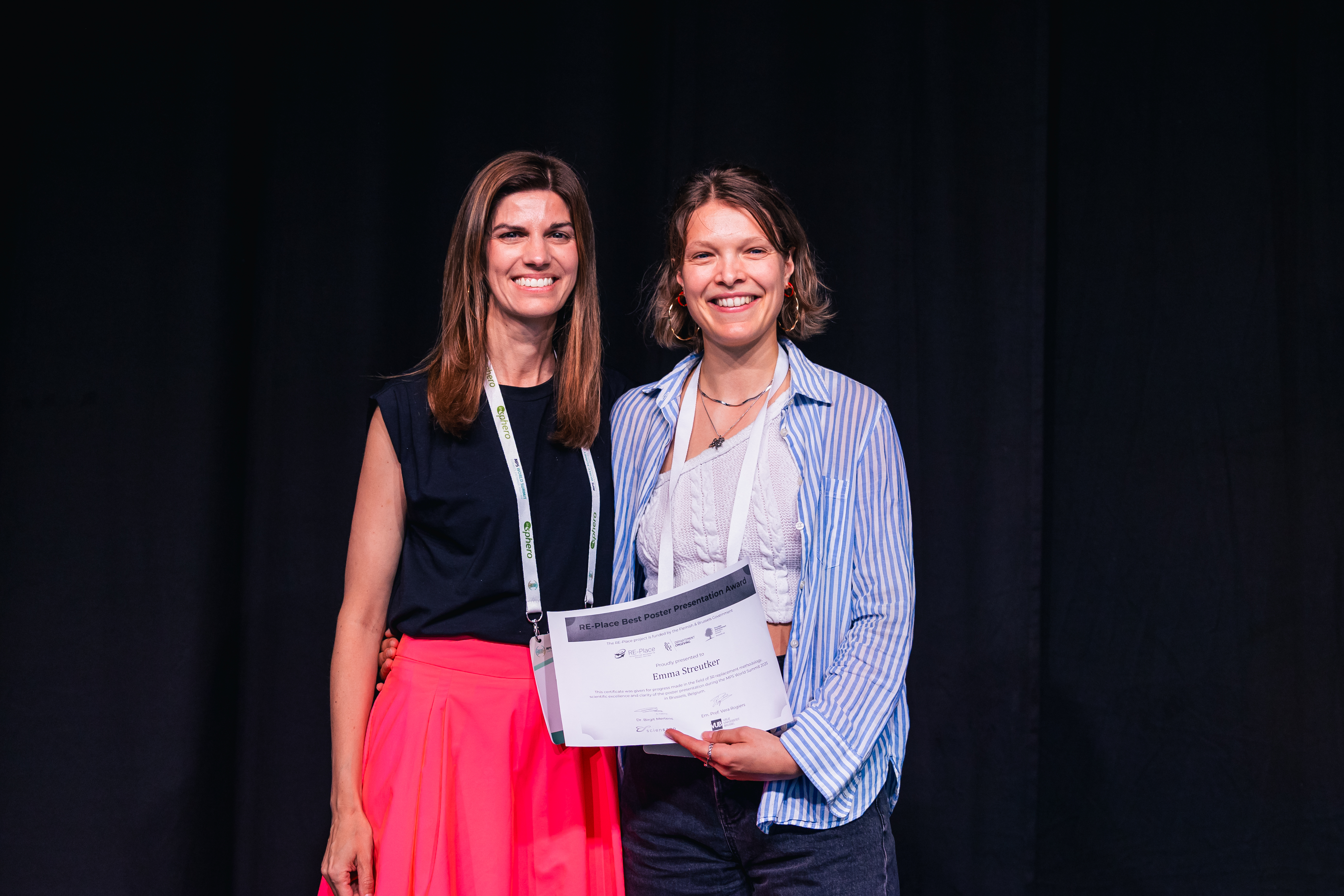
Emma Streutker wins RE-Place Best Poster Presentation Award at MPS 2025
During the Microphysiological Systems World Summit (MPS 2025) organised from 9 to 13 June 2025 in Brussels, the RE-Place team granted the RE-Place Best Poster Presentation award. The prize was open to PhD students and early career investigators presenting their research in the field of 3R replacement methodologies.
Emma Streutker, a PhD student at the Radboud Institute for Molecular Life Sciences, was awarded the prize for her poster titled “A fibrosis-on-chip model to study Systemic Sclerosis.” Her work was recognized for its scientific excellence, impactful results, and the clarity of her presentation.
RE-Place co-coordinator Dr. Birgit Mertens (Sciensano) presented the award during the conference's closing ceremony.
Congratulations to Emma!
Read her abstract:
Background: Systemic sclerosis (SSc) is a rare autoimmune disease characterized by inflammation, vasculopathy, and excessive fibrosis of connective tissues. Its exact pathophysiology is still incompletely understood, resulting in a lack of targeted therapies. Available animal and in vitro models have serious drawbacks relating to species differences and poor translational values. It is challenging to make an in vitro system encompassing an SSc phenotype that incorporates a relevant 3D environment and key cell types (fibroblasts, immune and endothelial cells) [1]. This project aims to address these issues by creating a novel in vitro fibrosis-on-chip model.
Methods: Polydimethylsiloxane (PDMS)-based organ-on-chip devices were produced using 3D-printed molds, bonded to glass slides, coated, and sterilized. Primary human dermal fibroblasts (HDF) were cultured in a 3D fibrin-collagen (2.5 mg/mL fibrinogen, 0.5 mg/mL collagen type l) matrix within the organ-on-chip device. Fibroblasts were stimulated with 10 ng/mL TGF-β for five days, and their profibrotic response investigated using confocal microscopy by staining transgelin, an actin cross-linking protein, and cell nuclei. A vascularized organ-on-chip was created by co-culturing HDFs and Human Umbilical Vein Endothelial Cells (HUVECs) 1:1 in a 3D fibrin (3 mg/mL) matrix for seven days. Vascular networks were assessed via confocal microscopy by staining CD31, an endothelial cell marker, and cell nuclei.
Results: HDF cultures stimulated with TGF-β showed an increased number of transgelin+ cells compared to unstimulated controls, as detected by immunofluorescent staining. Stimulated cells also showed distinct morphological changes, transitioning from a spindle-like, elongated shape to a larger, irregular form. To note, transgelin expression in non-stimulated controls was heterogenous within the HDF cultures. In the vascularized organ-on-chip model, co-culturing HDFs and HUVECs led to the spontaneous formation of 3D vascular networks. The vessel-like networks were found to have lumens as determined by 3D confocal imaging.
Conclusion: We have taken steps in developing a fibrosis-on-chip model to investigate SSc pathophysiology and evaluate therapies. Ongoing steps involve incorporating patient-derived fibroblasts and immune cells, evolving our model into a patient-specific SSc-on-chip model, with applications involving the study of SSc-induced vasculopathy.
[1] Streutker, E.M. et al. (2024). Adv. Healthcare Mater., 13, 303991. https://doi.org/10.1002/adhm.202303991


